MRF

Border Disease!!!!

I find it amazing that the media swallows the line that cases are centered in Texas, California and Florida.Some of the more fringe “media” outlets on the web are taking this, and suggesting it must be a disease bought over the border by illegal immigrants.

Let’s look at some infographics. Firstly from Morgellons.org, we have a little map that shows the states with the most cases:
txcaflmap.jpg
See, it’s Texas, Florida and California!

Now, here’s a more detailed map from “Unidentified Skin Parasite (a.k.a. “Morgellons Disease”) Database“. Darker states have more cases.
usa_morg_mapx.jpg

Finally, here’s one from Wikipedia:
usa_states_population_colorx.jpg

Notice the amazingly high degree of correlation between the maps! This proves without a doubt that cases are highest in CA, TX and FL!!

But wait, what’s Wikipedia doing supporting this nonsense?

Actually, the Wikipedia map in not a map of Morgellons incidence. It’s a map of population.

That’s right folk, there are more cases in CA, TX and FL, because MORE PEOPLE LIVE THERE!

The only reason those states have the most cases, is that they have the most people.

You might as well go around saying that the highest incidence of people is found in California, since it is, and it’s just as meaningful.

And CBS2 news in Los Angeles gets people all scared, saying that Los Angeles has the highest number of cases of Morgellons in the country. Could this possibly be related to the fact that (excepting New York) Los Angeles has MORE PEOPLE than any other city in the US?

Cause and Effect

Recently (April 2006) a member of the Morgellons Community, a young man, died of a drug overdose. He was one of several patients of Ginger Savely, a Texas Nurse Practitioner who offers diagnoses of Morgellons to those who seek it.

This young man was very intelligent and well-spoken. He was also a self-admitted drug abuser, taking a wide range of drugs including heroin. He was very open about this, and discussed his extensive drug use, and his related health problems, at great length on a large number of internet posts.

After he was “diagnosed” in early 2005, he apparently became increasing depressed and paranoid, believing that he was the subject of biological and electronic harassment.

His passing is very sad, but it is made sadder in that the Morgellons Community will memorialize him as a victim of Morgellons. I think that “Morgellons” probably did contribute a little to his fate, but not in the way that Savely and other would suggest.

It is very, very, sad.

We all have the same symptoms!

Morgellons folk say they all have the same symptoms.

  • You research the internet and so easily discover that Elliot’s disease, National United Skin Parasites Association, the Fiber Disease, and Morgellons are all one and the same. Ironically, all of the people with the exact same symptoms that you have, have been receiving the same faulty diagnosis

What are these symptoms? What does MRF have to say? Well, they have two pages, one on “symptoms, and one that proports to be a case definition. Symptoms:
http://morgellons.org/symptoms.htm
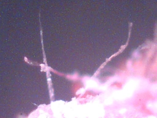
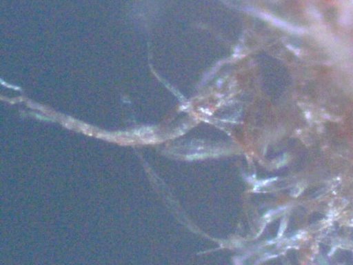
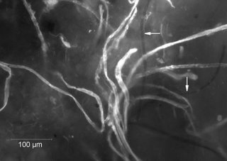
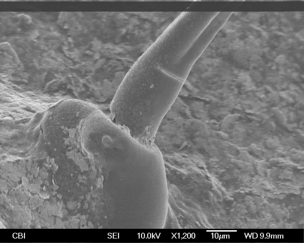
Most individuals with this disease report disturbing crawling, stinging, and biting sensations, as well as non-healing skin lesions, which are associated with highly unusual structures. These structures can be described as fiber-like or filamentous, and are the most striking feature of this disease. In addition, patients report the presence of seed-like granules and black speck-like material associated with their skin.[…] More significant than skin symptoms, in terms of the diminished quality of life of the individual with this illness, are symptoms unrelated to skin, to include Chronic Fatigue Syndrome (CFS), Fibromyalgia (ME), joint pain, and significant problems with concentration and memory.
Remarkably, not all people with this disease have overt skin lesions, as some individuals report intact skin. The troubling sensations and accompanying physical structures, are the consistent clues to this infectious process

http://morgellons.org/case.htm
[…]The following case definition of Morgellons disease has been developed by physicians on the medical advisory board of the Morgellons Research Foundation[…]

Now, I’m not going to post the entire thing here, as it’s quite long. But I’ll paraphrase the important parts:

  • Lesions may or may not be present, they might be a symptom of the disease, or the result of scratching. They might look like pimples, or hives, and may or may not contain pus.
  • Crawling sensations can occur anywhere on the body.
  • Fatigue is always present.
  • There will be behavioral effects that are diagnosed as psychiatric disorders (the implication is that they do not have these disorders, but the symptoms indicate Morgellons).
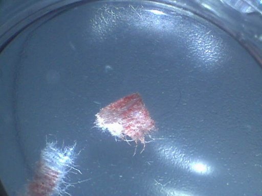
  • Fibers are reported in and on lesions and on the skin. Fibers may be any color size or shape. Granuals of any size or shape are found on the lesions and skin. Fuzzballs up to 3mm in size are found on lesions and skin.
  • Additonal symptoms include: vision changes, neurological changes, gastrointestinal changes, skin changes, Musculoskeletal changes.

Other than the fibers, what we have here is a catch-all of symptoms. Anyone who has some chronic condition cannot fail to have some of these symptoms. NOBODY has them all.

One can become a “Morgellons” sufferer simply by matching a few of your symptoms to the list, and then examining your skin for fibers – which everyone has on their skin.

What about those other conditions mentioned: Elliot’s Disease? Well, Elliot’s Disease research Library links to this article about “this disease”
http://explorepub.com/articles/omar_10_2.html#top
This gives a simpler definition:

  • It is characterized by neurological sensation of movement subcutaneously and/or in deeper tissues and cavities that is usually associated with mucoid cutaneous lesions from which one or more species of arthropods as well as unidentified fibers may be recovered.

Unfortunately, although this is a shorter definition, it’s actually even less precise than the Morgellons.org definition. We have a “sensation of movement” either on the skin or somewhere on the body, usually with lesions but not always, and either with or without arthropods and with or without fibers. Based on that definition – I’ve got it, since my scalp itches a bit right now. Yours probably does too.

Moving on, NUSPA has a nice page of symptoms:

http://www.skinparasites.com/id12.html

They focus on a very broad range of symptoms of infestation by a parasite, insomnia and the specks and fibers. Nowheres does it mention fatigue or any neurological complain. This is obviously very different to Morgellons.

Then there is “The Fiber Disease” – this seems to be a term popularized by the biology-online forum thread.
http://www.biology-online.org/biology-forum/about1958.html
Unfortunately, they don’t list the symptoms anywhere I could find. But individual posters report a very wide range of symtoms.

Okay, finally my point:

Not everyone has the same thing.

There are so many different symptoms, it’s impossible to describe this as “a disease” or even “a syndrome”. Everyone has different symptoms, everyone responds differently to treatments. The term “Morgellons” was invented to describe a child’s skin problems, which he is now cured of, and which have no relation to the symptoms described by the vast majority of sufferers.

Morgellons in California

Why is there so much Morgellons in California?

Morgellons.org states:

Note: Although California represents 12% of the US population, 26% of all who have registered with the Morgellons Research Foundation reside in California

Now it’s tempting to say things like “California is full of dirty hippy druggies!”, but that’s not really true. Besides, most of the Morgellons sufferers are nice middle-aged white women.

So, why this disproportionate number of Morgellons sufferers from California?

Note what the above is measuring – it’s a percentage of all who have registered with the Morgellons Research Foundation. i.e, over the internet.

How do people find out about Morgellons – on the internet.

How do people share their photos of fibers – on the internet.

How do people form communities to share stories of their illness?

The internet – a common factor in Morgellons suffers.

Morgellons is an interesting syndrome because of the visual aspect – fibers, and hence photos of fibers. Lots of sites have many many photos of fibers. These sites really need a broadband internet connection (DSL or Cable Internet).

In June 2004, California had 13 broadband lines per 100 persons, and the rest of the US had 9.

http://www.m9systems.com/broad_dep_2.html

Could it be that there is more Morgellons, because there is more broadband?

These simple figures don’t tell the whole story either – California’s population is clustered into cities. The Morgellonites in CA also cluster around the four major population centers. In those population centers, the access to broadband internet is unparalleled through the United States. People in Urban California have the best availability to broadband internet in the country.

If Morgellons is a phenomenon fueled by the internet, then obviously there would be more cases in California.

California is a unique state in many ways – perhaps there are some other factors?

The far side of Morgellons meets the near side.

In the Morgellons community, there are several folk who think that their problems are due to some kind of genetically modified organism, typified by this post from TamTam

Fibers are stray target of experimental biology.
In particular experimental micro biology class 3 (C3)

That cellulose type fibers move on its own is because it are micro organisms that go dormant (pseudo apoptotic) In particular micro organisms that breath and become static because of low metabolism.
During formation they show nerve cell type behavior.

Nerve cell type behavior they will show in particular when they unite as a fiber most resembling a shaft that is related to the mold of the cyanobacterium.

This dominant type gene expression in the form of a semi autonomous and sensing fiber is in fact a break away group cell.
A variant form.

Its character is inbetween plant(cellulose) and insect (chitin)cell.

Single cell micro organisms with an aquired property called quorum sensing will make the organisms act multi cellular.

Quorum sensing is a stem cell like property.
Quorum sensing micro organisms unite hence differentiate during formation of a multi cellular organism.

Hence multi lineage differentiation synonymous with stem cell.
————————————————————————————–
Fast dividing tissue forms a base(culture medium) for this organism.
This type target can only be kept alive and cultered in test animal.
————————————————————————————–

Fibers show inbetween behavior like “plant cell that think they are insect cell”

The agent its intermediate stage resembles a protoplasm.
A transparent cloud with basic properties preserved.

All elements are quorum sensing
All elements differentiate

check: chitin / cellulose wound dressing

TamTam writes in an unusual way. Declarative sentences with little explanation. This reminds me of the way AIs are portrayed in Science Fiction. Or perhaps someone with a brain wired a particular way – maybe Asbergers (not that there’s anything wrong with that, Bill Gates has often been diagnosed with Asbergers).

TamTam is probably responsible for this video:

http://www.silentsuperbug.com/

Which shows text similar to the above, overlaid on footage of some microscopic video.

Interstingly, mainstream Morgellons hits fringe Morgellons when Randy Wymore analyses this video on mogellon.org
http://morgellons.org/rwupdate.html
He basically says “there is no science here”, but uses several more word, trying to be kind.

Objects in lesion on child’s lip look like Kleenex fibers

Morgellons.org has, for a long time (since 2002), shown two pictures titled:

Objects emerging from lesion on child’s lip at 200x

The two photos were probably taken with a QX3 microscope, and have an image size of 512×384 pixels (standard size for QX3 and QX5 microscopes) and they exhibit the characteristic lighting of QX microscopes.

Here is one of the photos from Morgellons.org:

morgellons_drew2-200x.jpg

Here is a photo I took on a QX5 at 200x

blood-kleenex-200x-2.jpg

Notice the similarities. The size of the “structures” are almost identical. They exhibit a characteristic “flattening” in places. They have similar lengths, thickness and variety.

What are they? Kleenex tissue, stained with blood.

Here’s the same thing at 10x

blood-kleenex-10x.jpg

The only difference here is in the color, which can be accounted for by the lighting, and the amount of blood soaked into the sample. Mine just had a tiny bit of blood on it, so the fibers are not fully soaked.

So, the fibers that were found in a lesion on a child lip look exactly like Kleenex soaked in blood. Does it seem at all unlikely that if you (or your child) has some seeping lesions on their lip, then they might at some time dab them with a Kleenex, perhaps leaving a few fibers?

Morgellons.org also says the fibers “have tentatively been identified as cellulose”. What is Kleenex made from? Cellulose (refined from wood pulp).

While this does not explain the dark fibers in other photos, it’s an obvious and simple explanation for the “Objects emerging from a lesion”, and Morgellons.org should no longer be claiming that these are somehow unusual, unidentified, hyphae-like structures.

They look exactly like Kleenex fibers, soaked in blood. That’s probably what they are.

An early exchange

Back in July 2002, Mary Leitao posted a request for help on the medhelp.org buletin board, and entered into a brief discussion with Dr Rockoff (a dermatologist). Dr Rockoff was very polite. After a few back and forth exchanges the thread devolved into a “me too!” state.

Dr Rockoff’s responses are very reasonable, and should be a model for similar exchanges.

http://www.medhelp.org/perl6/dermatology/messages/31706a.html

Hypha? Hair?

Consider the photos here:
http://www.morgellons.org/symptoms.html


* “These structures can be described as fiber-like or filamentous”
* “The unknown fibers associated with skin lesions can be described as coenocytic (aseptate), smooth-walled, branching, filamentous objects. The fibers have been analyzed by FTIR (Fourier Transform Infrared Spectroscopy) and have tentatively been identified as cellulose.”
* “The fibers are clearly hyphae-like structures, and yet, do not fall within the description of known hyphae or pseudohyphae

So can the items in the photos be described as “coenocytic” or “hyphae-like”? The two photos are titled “Objects emerging from lesion on child’s lip at 200X”. Now given this “200X”, how long does that make the “objects”? We can’t really say, as the images themselves could be scaled to any size. But approximately, the “fibers” on my screen are 40mm long, and 2mm thick. This makes them 0.2mm long, and 0.01mm thick. (200 microns by 10 microns, where 1 micron=0.001mm). This puts them approximately in the same scale as hyphae (the living, growing parts of fungi, tiny tubes filled with cytoplasm and nuclie).

On an old page at morgellons.org, that is still up, but seemingly not linked to, it says:
“The fibers are clearly hyphae-like structures, and yet, due their immense size (150 microns wide), they do not fall within the description of known hyphae or pseudohyphae. “
Huh? Even if measuring off 1:1 the screen, then at 200x magnification, 150 microns would appear as 30mm wide (about an inch). Plainly the objects are not 150, and are more like 15 microns thick. Either that or the photos are labled incorrectly. However, the SEM photos clearly show “fibers” that are about 15 microns.

Hypha can be both septate and aseptate (Coenocytic). Septate hyphae are divided by cross section segments called septa. Aseptate hyphae are not. Detrmining if a structure is septate or aseptate is difficult with the images provided, as the most of the structures are out of focus. The clearest structure (in the first image) actually shows some evidence of being septate, rather than aseptate. The picture appears to have been taken with reflective light (from above), and a better determination could be made with transmissive light (from below). Additionally, the specimen should be mounted on a slide and stained to enhance contrast.

Additional stained images are given here:
http://www.morgellons.org/images.html

However, this photo does not resemble the first photos at all, clearly they are different structures, much longer, and smoother.

Then we have some SEM (Scanning Electron Microscope) photos. These photos reveal surface features.

Here we have a scale, showing the object is about 15 microns thick. Human hair is generally described as “60-100 microns in diameter”, which would seem to eliminate that as a candidate. However, that is the diameter of full grown head hair (a “terminal” hair). Finer new grown body hair will emerge with an initial diameter that is much smaller, children are covered in “vellus” hair, which has a diameter of under 30 microns.

Going back to the first photo, since the “structures” here are from a lesion on the lip of a child”, and the lesions clearly extend beyond the actual lip surface, then is it not reasonably to think they might be vellus hairs, coated in some viscous liquid (blood, puss or sebum)?

In this photo of the site from which the specimen was obtained:

You can clearly see what look like vellus hair in the lower portions of the image

Use of words

Morgellons proponents sometimes use technical language in describing their findings. I feel this clouds the meaning somewhat.

consider:

http://www.morgellons.org/symptoms.html
The unknown fibers associated with skin lesions can be described as coenocytic (aseptate), smooth-walled, branching, filamentous objects. The fibers have been analyzed by FTIR (Fourier Transform Infrared Spectroscopy) and have tentatively been identified as cellulose.”

What does this mean?

coenocytic – I like how this is “explained” as being “aseptate”, as if this will somehow help the reader understand. Of course, you can look these words up, on the internet.

Coenocytic basically means cells with multiple nuclei. Aseptate can mean the same thing, but more literally means lacking the cell walls (septate) between nuclei. This type of cellular growth is typical of the threads of many fungi and some algae. See:
http://www.botany.hawaii.edu/faculty/wong/BOT135/Lect03_b.htm

“Smooth walled” seems a little odd, as the example photos at the top of the “symptoms” page do not look smooth walled at all. The next set of photos show some very thin black fibers, that appear at first glance to be smooth walled (but you can’t really tell), which surround a much thicker white fuzzy object. Where did “smooth walled” come from?

“FTIR” is your standard chemical spectography thing. You measure the absoption of various wavelengths of light, and that gives you graphs you can compare against known substances to see if it matches. A tentative identification as “cellulose” is not surprising. Cellulose is the most abundant form of living biomass on the planet. Cotton, for example, is composed largely of cellulose (91% cellulose, 8% water, 1% other).
http://www.tis-gdv.de/tis_e/ware/fasern/baumwoll/baumwoll.htm. Other cellulose fibers are linen, ramie, rayon, tencel and lyocell all of which are almost purely cellulose. Paper and paper tissues are also largely comprised of cellulose fibers.